hCG: Why Human Chorionic Gonadotropin Is a Glycoprotein, Not a Peptide
hCG is often called a peptide, but it is a glycosylated heterodimer. Its structure explains IU labeling, analytical methods and storage needs.
 EMElena Marsh, PhDSenior Research Analyst, Analytical Chemistry~6 min read
 CompoundsPhoto: Kjaergaard / Wikimedia Commons
Search queries for “hcg peptide” are common, and research suppliers often list human chorionic gonadotropin beside short synthetic peptides. Structurally, though, hCG belongs to a different class of molecule. It is a heterodimeric glycoprotein of roughly 37 kDa, assembled from two non-covalently associated subunits and carrying a substantial load of carbohydrate. Those features govern almost everything a laboratory needs to know about it: how it engages its receptor, why its activity is reported in International Units instead of milligrams, which analytical methods can characterize it, and how it should be stored.
This profile stays at the level of protein chemistry, receptor pharmacology and analytical practice. Research-grade hCG is for laboratory research use only; not for human or veterinary use.
The structure of hCG: two subunits, one hormone
hCG is a member of the glycoprotein hormone family, which also includes luteinizing hormone (LH), follicle-stimulating hormone (FSH) and thyroid-stimulating hormone (TSH). All four are heterodimers built from a common alpha subunit and a hormone-specific beta subunit. Neither subunit has meaningful receptor activity on its own; biological activity depends on the correctly folded, non-covalently associated αβ dimer.
The common alpha subunit
The alpha subunit is a 92-residue polypeptide whose amino acid sequence is shared by all members of the family within a species. It contains five disulfide bonds and carries two N-linked oligosaccharides.
The beta subunit and its C-terminal peptide
Receptor specificity comes from the 145-residue beta subunit. hCGβ shares most of its sequence with LHβ, which is why the two hormones act at the same receptor. Its distinguishing feature is a C-terminal extension of roughly 30 residues, usually called the C-terminal peptide (CTP), which is rich in serine and proline and carries four O-linked glycans. Together with two N-linked glycans on the beta chain, that gives eight glycosylation sites per dimer, and carbohydrate accounts for roughly a quarter to a third of the molecule’s mass.
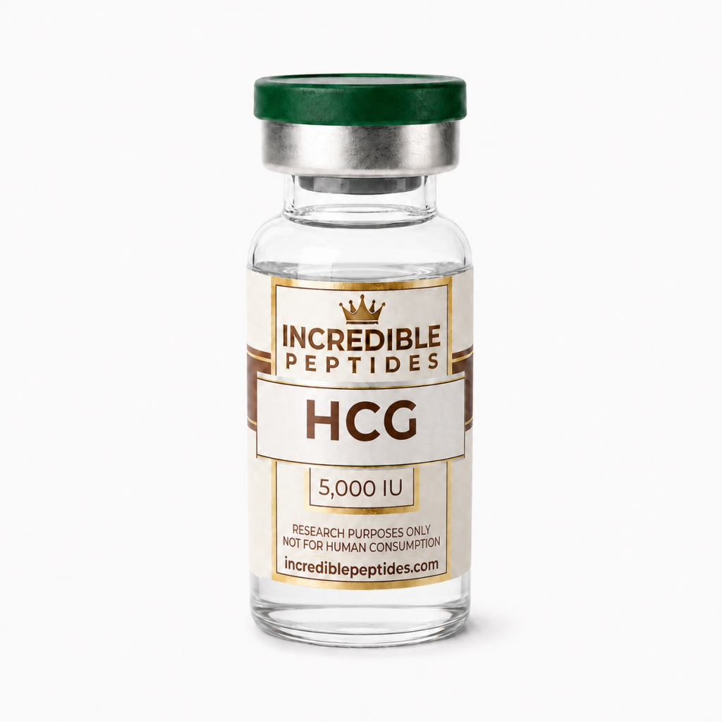
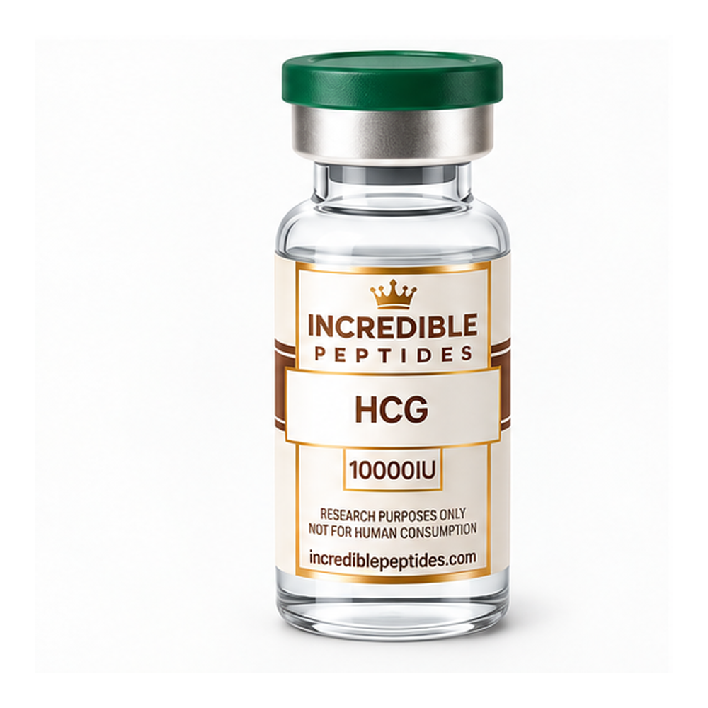
The crystal structure published in 1994 showed that each subunit adopts a cystine-knot fold, the architecture also found in growth factors such as nerve growth factor and transforming growth factor-β. A segment of the beta subunit, often described as a “seatbelt”, wraps around the alpha subunit and is latched by a disulfide bond, which stabilizes the heterodimer. This structure is also why a lyophilized vial such as the HCG 5000 IU research material is labeled by biological activity rather than by peptide mass.
Peptide or protein? Why the classification matters
Most chemists reserve “peptide” for chains of up to about 50 residues that can be made by solid-phase synthesis and characterized largely by sequence and mass. hCG fails each test. Its 237 residues are distributed across two chains, its function depends on correct disulfide pairing and subunit assembly, and its glycans are heterogeneous. Cellular glycosylation also yields a mixture of glycoforms differing in branching and sialic acid content.
The practical consequence is that hCG has to be produced biologically, either purified from a natural source or expressed recombinantly in a mammalian cell line capable of complex glycosylation. The broader distinction is covered in our article on peptides vs proteins.
The LH/CG receptor and its signaling
hCG and LH share a single receptor, the luteinizing hormone/choriogonadotropin receptor (LHCGR). It is a class A G protein-coupled receptor with an unusually large extracellular domain built from leucine-rich repeats, which forms the principal binding surface for the hormone. A hinge region links it to the seven-transmembrane core.
In cell systems expressing LHCGR, the dominant response is coupling to Gs, activation of adenylyl cyclase and accumulation of cyclic AMP, followed by protein kinase A signaling. At high receptor occupancy, coupling to Gq/11 and phospholipase C has also been described. Older in-vitro work found that chemically or enzymatically deglycosylated hCG still binds the receptor yet produces a much weaker cAMP response, an early indication that the carbohydrate is not inert decoration. These readouts underpin the cell-based bioassays used to compare preparations, typically cAMP accumulation in a recombinant cell line or steroid output from cultured Leydig cells. Background on these formats is available in our guide to cell signaling assays.
Why hCG activity is expressed in International Units
Mass is a poor predictor of glycoprotein activity. Two lots with identical protein content can differ in glycoform distribution, in the proportion of free subunits and in the fraction of correctly assembled dimer. Activity therefore has to be measured directly and reported relative to a reference.
That reference is a WHO International Standard for chorionic gonadotropin: a carefully characterized lyophilized preparation, held by the National Institute for Biological Standards and Control (NIBSC), to which a defined number of International Units is assigned by collaborative study. Each replacement standard is calibrated against its predecessor. An IU is consequently a defined quantity of activity in a reference bioassay rather than a fixed mass, and the conversion between IU and micrograms varies with the preparation and the method.
Historically, potency was assigned with in-vivo bioassays in rodents, such as organ-mass responses of androgen-dependent tissues in immature rats. Current practice leans increasingly on in-vitro and physicochemical methods, but the IU remains the reporting convention. That is why research vials come in activity sizes, for example a 10,000 IU lyophilized hCG vial, and why the matching certificate of analysis should state the reference standard and assay against which content was assigned.
Analytical characterization of a glycoprotein
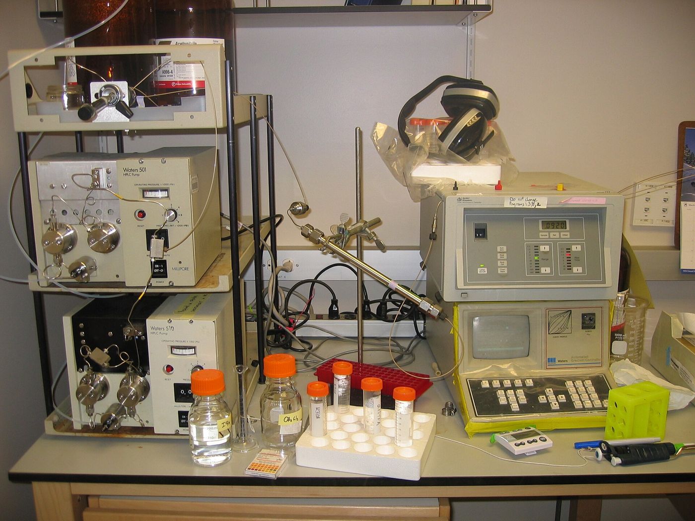
A single HPLC purity figure, adequate for many short peptides, says relatively little about hCG. Characterization instead combines orthogonal methods, each answering a narrower question. The table summarizes the main ones.
| Method | What it shows | Main limitation |
|---|---|---|
| SDS-PAGE (reducing and non-reducing) | Subunit and dimer pattern, gross protein contaminants | Glycan heterogeneity gives broad, diffuse bands |
| Size-exclusion chromatography | Aggregates, intact dimer versus free subunits | Limited resolution between similar species |
| Reversed-phase HPLC | Subunit variants and process-related impurities | Denaturing conditions dissociate the dimer |
| Mass spectrometry with peptide mapping | Sequence confirmation, glycosylation site occupancy, glycan composition | Intact-mass spectra are complex because of glycoform spread |
| Immunoassay | Dimer- or subunit-specific epitope content | Does not measure receptor activity |
| Cell-based bioassay | Activity in IU against a reference standard | Higher variability than physicochemical methods |
ICH Q6B frames the logic well: identity, purity, heterogeneity and potency are separate attributes needing separate evidence.
Handling and storage considerations
Because activity depends on the non-covalent dimer, conditions that separate the subunits destroy function even when no covalent bond breaks. Low pH, elevated temperature, chaotropic agents and organic solvents can all dissociate alpha from beta, and free subunits do not readily reassemble in dilute solution. Sialic acids are also acid-labile, so prolonged acidic exposure can shift the glycoform profile.
Lyophilized glycoprotein is generally far more stable than the same material in solution. For in-vitro work, standard protein-laboratory practice applies: prepare single-use aliquots in a suitable buffer, avoid repeated freeze-thaw cycles, and allow for adsorption of dilute protein to plastic and glass, often by including a carrier protein where the assay permits. Lot documentation should come first, however; the hCG research vial listing and its COA are where storage conditions and assigned activity should be confirmed before material enters an experiment.
Key points for researchers
hCG is best understood as a glycoprotein hormone rather than a peptide: a cystine-knot heterodimer of about 37 kDa whose carbohydrate makes up a large share of its mass and shapes its receptor signaling. That structure explains its biological production, its IU labeling against a WHO standard and its reliance on orthogonal analytics. For any lot, the most useful questions are which reference standard its IU content was assigned against, how dimer integrity was assessed and how the material should be stored.
References
- Lapthorn AJ, Harris DC, Littlejohn A, et al. Crystal structure of human chorionic gonadotropin. Nature, 1994.
- Pierce JG, Parsons TF. Glycoprotein hormones: structure and function. Annual Review of Biochemistry, 1981.
- Ascoli M, Fanelli F, Segaloff DL. The lutropin/choriogonadotropin receptor, a 2002 perspective. Endocrine Reviews, 2002.
- International Council for Harmonisation. ICH Q6B: Specifications: Test Procedures and Acceptance Criteria for Biotechnological/Biological Products.
- WHO Expert Committee on Biological Standardization. Technical Report Series (reports establishing International Standards for chorionic gonadotropin).
Research Products in This Article
Educational content for research use only. Not for human consumption. Nothing here is medical advice or a claim about safety or efficacy.
 DO