Melanotan II (MT-2): Structure and Melanocortin Receptor Pharmacology
How melanotan II was designed from alpha-MSH, why its lactam ring matters, and how it behaves at melanocortin receptors in cell-based assays.
 DODaniel Okafor, MSResearch Analyst, Molecular Biology~5 min read
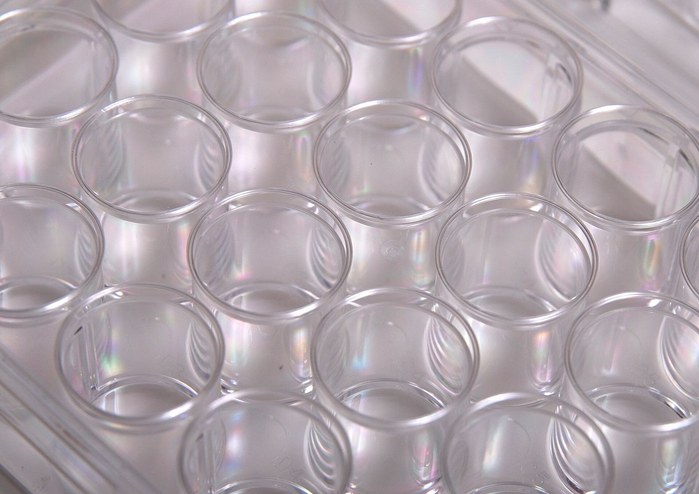 CompoundsPhoto: Lilly_M / Wikimedia Commons, CC BY-SA 3.0
Melanotan II, often abbreviated MT-2 or MT-II, is a synthetic cyclic analogue of alpha-melanocyte-stimulating hormone (alpha-MSH) and one of the most widely used tool compounds in melanocortin receptor research. Interest in the mt2 peptide comes from its compact lactam-bridged structure, its high potency in cell-based assays and its broad agonist activity across the melanocortin receptor family. It also served as the starting scaffold for several important analogues, including the antagonist SHU9119 and the closely related free-acid peptide PT-141.
This article describes the sequence and design history of melanotan II, its receptor pharmacology at MC1R through MC5R, its use in melanocyte and melanoma cell-culture models, and the practical points that matter when sourcing and characterising a melanotan II research peptide. Like all compounds discussed here, it is intended for laboratory research use only; not for human or veterinary use.
From alpha-MSH to a constrained analogue
Alpha-MSH is a 13-residue peptide, Ac-Ser-Tyr-Ser-Met-Glu-His-Phe-Arg-Trp-Gly-Lys-Pro-Val-NH2, produced by processing of pro-opiomelanocortin (POMC). Structure-activity work in the 1970s and 1980s established that the central His-Phe-Arg-Trp segment carries most of the information needed for receptor activation, while the flanking residues mainly influence potency and stability.
Two substitutions proved especially productive. Replacing Met4 with norleucine removed an oxidation-sensitive thioether, and replacing L-Phe7 with D-Phe greatly increased potency and resistance to enzymatic breakdown. The resulting linear analogue, [Nle4, D-Phe7]-alpha-MSH (NDP-MSH, sometimes called melanotan I), was described by Sawyer, Hruby, Hadley and colleagues in 1980 and remains a standard high-affinity reference ligand. Radioiodinated NDP-MSH is still the most common tracer in melanocortin binding assays.
Melanotan II was the next step. Working at the University of Arizona, Hruby’s group shortened the sequence to the 4-10 region and introduced a side-chain-to-side-chain lactam bridge, using molecular dynamics simulations to choose ring positions that would stabilise a receptor-compatible turn. The resulting cyclic peptides, reported in 1989, were markedly more potent than the linear parent in the amphibian melanophore bioassays used at the time.
Sequence and physicochemical properties
Melanotan II has the sequence Ac-Nle-cyclo[Asp-His-D-Phe-Arg-Trp-Lys]-NH2. It is a heptapeptide with an acetylated N-terminus, a C-terminal carboxamide and an amide bond between the beta-carboxyl of Asp and the epsilon-amine of Lys. The molecular formula is C50H69N15O9 and the average molecular weight is approximately 1024.2 g/mol.
The ring encloses the His-D-Phe-Arg-Trp pharmacophore and constrains it in a beta-turn-like geometry centred on D-Phe and Arg. Arginine and histidine contribute positive charge, so the peptide is basic overall, and the tryptophan indole gives strong UV absorbance near 280 nm alongside the peptide-bond absorbance at 214 to 220 nm used for HPLC purity assessment. The lactam bridge and D-amino acid together make melanotan II considerably more resistant to proteolysis in serum and tissue preparations than native alpha-MSH, which is part of why it became a favoured in vitro and preclinical tool.
Melanotan II and PT-141
PT-141 (bremelanotide) shares the identical cyclic core but ends in a free carboxylic acid rather than an amide. The two differ by roughly 1 Da, so analytical methods must be able to distinguish them. Our profile of PT-141 and its cyclic melanocortin structure covers that compound separately.
Receptor pharmacology
The five melanocortin receptors are class A GPCRs that couple mainly through Gs to adenylyl cyclase, raising intracellular cAMP. MC2R is selective for ACTH and needs the accessory protein MRAP to reach the cell surface, so it is not a relevant target for MSH analogues. In cell lines expressing the other four subtypes, melanotan II acts as a potent, largely non-selective full agonist at MC1R, MC3R, MC4R and MC5R, with reported EC50 values typically in the sub-nanomolar to low nanomolar range. Exact values depend heavily on the expression system and readout and should be compared only within a single study.
Because it activates several subtypes, melanotan II is not a good tool for attributing a cellular response to one receptor. Researchers usually combine it with subtype-selective antagonists, receptor knockdown, or cell lines that express a single receptor. When an analogue is needed for direct comparison, the free-acid PT-141 research peptide is a natural partner, since it isolates the effect of the C-terminal modification on an otherwise identical scaffold.
SHU9119: one substitution, opposite effect
Replacing D-Phe in melanotan II with D-2′-naphthylalanine produces SHU9119, reported by Hruby and co-workers in 1995. SHU9119 is a potent antagonist at MC3R and MC4R yet remains an agonist at MC1R and MC5R. This pair is a textbook example of how the size of a single aromatic side chain inside a constrained ring can switch efficacy at some receptors while leaving others unchanged, a theme discussed further in our article on peptide structure-activity relationships.
Melanocyte and melanoma cell models
MC1R is the predominant melanocortin receptor on melanocytes, and much of the in vitro literature on melanotan II concerns pigment-cell biology. In cultured melanocytes and in murine B16 melanoma cells, MC1R activation raises cAMP, activates protein kinase A and the transcription factor CREB, and increases expression of the microphthalmia-associated transcription factor (MITF). MITF in turn drives transcription of melanogenic enzymes such as tyrosinase, tyrosinase-related protein 1 and dopachrome tautomerase.
Typical readouts include intracellular cAMP, tyrosinase enzyme activity measured by L-DOPA oxidation, melanin content measured spectrophotometrically after cell lysis in alkali, and qPCR or immunoblotting for MITF and its targets. Experiments are usually run alongside alpha-MSH or NDP-MSH as reference agonists and, where receptor dependence must be shown, with MC1R-deficient cells or an antagonist. These are cell-culture findings, and the translation of any such observation beyond the culture dish is outside the scope of this article.
Sourcing and handling for assay work
Reliable pharmacology starts with confirmed identity. Mass spectrometry should match the amidated structure at about 1024.2 Da, and reversed-phase HPLC should show a single main peak resolved from potential impurities such as deletion sequences, uncyclised linear precursor, oxidised tryptophan species and any free-acid analogue. When purchasing a lyophilised MT-2 reference standard, check that the lot-specific certificate of analysis reports both identity and purity and that the counter-ion is stated, since acetate or trifluoroacetate content affects the net peptide amount weighed. Our guide to peptide mass spectrometry explains how to read those data.
Lyophilised material is best kept frozen, dry and protected from light because of the oxidation-prone tryptophan and histidine residues. Solutions for assays should be freshly prepared or aliquoted and frozen once, with the buffer and storage time recorded.
Melanotan II as a research tool
Melanotan II illustrates how a rational, structure-guided redesign of a native hormone can produce a stable, potent and synthetically tractable probe. Its non-selective agonism makes it valuable as a positive control across the melanocortin family and as a parent scaffold for analogues, while its well-defined chemistry makes it a useful case study in how cyclisation and D-amino acids change peptide behaviour.
References
- Sawyer, T. K. et al. 4-Norleucine, 7-D-phenylalanine-alpha-melanocyte-stimulating hormone: a highly potent alpha-melanotropin with ultralong biological activity. Proceedings of the National Academy of Sciences USA, 1980.
- Al-Obeidi, F., Castrucci, A. M., Hadley, M. E. and Hruby, V. J. Potent and prolonged acting cyclic lactam analogues of alpha-melanotropin: design based on molecular dynamics. Journal of Medicinal Chemistry, 1989.
- Hruby, V. J. et al. Cyclic lactam alpha-melanotropin analogues of Ac-Nle4-cyclo[Asp5,D-Phe7,Lys10]alpha-melanocyte-stimulating hormone-(4-10)-NH2 with bulky aromatic amino acids at position 7 show high antagonist potency and selectivity at specific melanocortin receptors. Journal of Medicinal Chemistry, 1995.
- Mountjoy, K. G., Robbins, L. S., Mortrud, M. T. and Cone, R. D. The cloning of a family of genes that encode the melanocortin receptors. Science, 1992.
- Cone, R. D. Studies on the physiological functions of the melanocortin system. Endocrine Reviews, 2006.
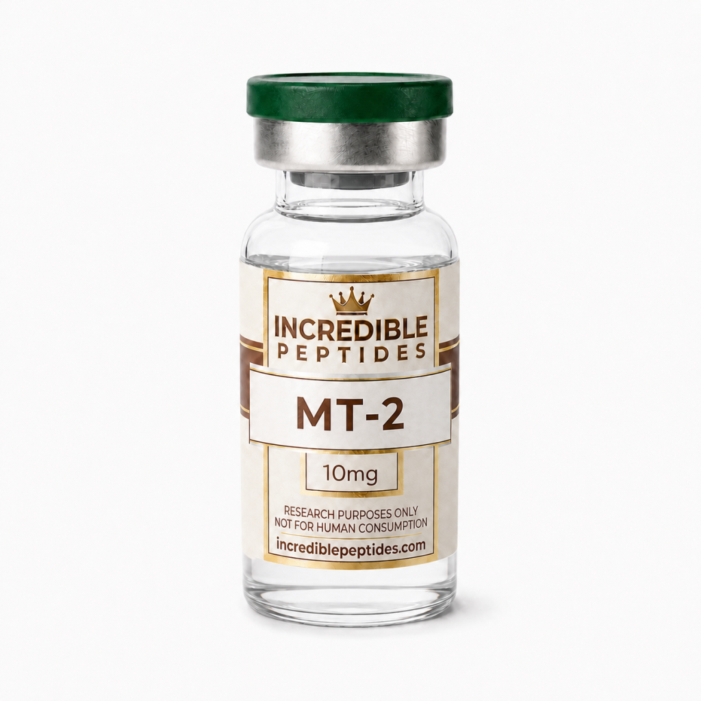
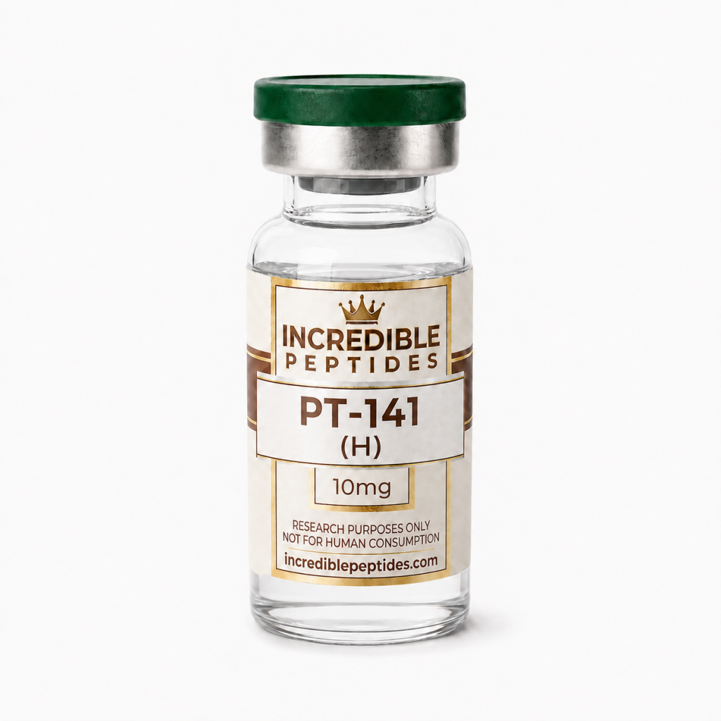
Research Products in This Article
Educational content for research use only. Not for human consumption. Nothing here is medical advice or a claim about safety or efficacy.