Glutathione: The γ-Glutamyl Tripeptide Antioxidant in Research
Glutathione is a γ-linked tripeptide whose cysteine thiol drives its redox chemistry, assay behavior and instability in solution.
 EMElena Marsh, PhDSenior Research Analyst, Analytical Chemistry~5 min read
 CompoundsPhoto: Millrock / Wikimedia Commons, Public domain
Glutathione is the most common small thiol in most animal cells. A thiol is a compound with a sulfur-hydrogen group. Glutathione is also one of the most often measured molecules in redox biology, the study of how cells gain and lose electrons. In a strict chemical sense, it is an unusual glutathione peptide. It is a tripeptide made of three amino acids: glutamate, cysteine and glycine. But the first bond forms through the side-chain carboxyl of glutamate, not its alpha-carboxyl. That one detail, plus the reactive cysteine thiol, explains most of how it behaves. This holds in cells, in assay buffers and in a storage vial.
This profile covers glutathione’s chemistry and its enzymes. It also covers its roles in cell-culture and assay work. Last, it looks at the practical problem of keeping the reduced form reduced. Research-grade material is for laboratory research use only; not for human or veterinary use.
Structure: a γ-glutamyl tripeptide
Reduced glutathione (GSH) is γ-L-glutamyl-L-cysteinylglycine. Its formula is C10H17N3O6S, and its molecular weight is about 307.3 g/mol. Its oxidized partner is glutathione disulfide (GSSG). GSSG is two GSH molecules joined by a disulfide bond. It weighs about 612.6 g/mol.
Why the gamma link matters
In a normal peptide, each residue is joined through its alpha-carboxyl group. In glutathione, glutamate is joined to cysteine through its γ-carboxyl, the side-chain acid. Most peptidases, the enzymes that cut peptides, do not see this bond. That is one reason GSH lasts at millimolar levels inside cells. Instead, its breakdown starts with γ-glutamyl transpeptidase. This enzyme sits on the outer surface of many cell types. It moves the γ-glutamyl group to an acceptor and frees cysteinylglycine. The wider rules of amide-bond formation are covered in our article on peptide bonds and amino acid sequences.
The cysteine thiol
The chemistry of glutathione is mostly the chemistry of its cysteine sulfhydryl group. The thiol pKa is around 9. So at neutral pH, only a small share exists as the more reactive thiolate anion. That share sets the rate of disulfide exchange, metal binding and reaction with electrophiles. It also rises sharply as pH goes up. So it is best to think of a freeze-dried reduced glutathione (GSH) research material as a reactive thiol reagent as much as a peptide.
How cells make and break down glutathione
Cells build glutathione in two steps. Both steps use ATP, and neither one involves the ribosome. First, glutamate-cysteine ligase joins the γ-carboxyl of glutamate to cysteine. Then glutathione synthetase adds glycine. The first step is the slow, rate-limiting one, and GSH itself holds it back through feedback. This pathway is the basis of a classic lab tool. Buthionine sulfoximine (BSO) is a selective blocker of glutamate-cysteine ligase. It drains cellular glutathione in culture and in animal models. It is widely used to ask whether a response depends on the glutathione pool.
Enzymes make glutathione from free amino acids. Because of this, it sits at an odd border. It is listed in the research peptides category because, in chemical terms, it is a tripeptide. Yet its biology looks more like that of a metabolite. Its synthesis, export and breakdown form what Meister described as the γ-glutamyl cycle.
The GSH/GSSG redox couple
Glutathione’s main role is as a source of electrons, or a reducing equivalent. Glutathione peroxidases, many of them selenoenzymes, reduce hydrogen peroxide and organic hydroperoxides. They do this by oxidizing two molecules of GSH to one of GSSG. Glutathione reductase then makes GSH again from GSSG using NADPH. This links the glutathione pool to the pentose phosphate pathway. Glutathione S-transferases use GSH as a cosubstrate to attach it to electrophilic compounds. Glutaredoxins use it to reduce protein disulfides and mixed disulfides.
In resting cultured cells, total glutathione is usually in the low-millimolar range. The GSH:GSSG ratio is high. Two GSH molecules are used up for each GSSG formed. So the reduction potential of the couple depends on total amount as well as on the ratio. Schafer and Buettner stressed this point. If you report the ratio alone, without absolute amounts, you can hide real differences in redox state between samples. Shifts toward GSSG are a common readout of oxidative stress in cell models. Reversible S-glutathionylation of protein cysteines is also studied as a control step in its own right.
Glutathione in cell culture and assay work
In the lab, glutathione plays three distinct roles. First, it is a reagent. GSH and GSSG are mixed in refolding buffers to help disulfide bonds form correctly. GSH is the cosubstrate in glutathione S-transferase activity assays. It is also the agent used to wash GST-tagged fusion proteins off their column. Second, it is a measured endpoint. It is quantified in cell lysates and in tissue extracts from animal models. Third, it is a test variable. It is added to or depleted from cultures to test redox dependence.
Measuring GSH and GSSG
Ellman’s reagent (DTNB) reacts with free thiols and releases a yellow dye. But it measures total sulfhydryl content, not glutathione alone. Tietze described an enzyme recycling method. It couples DTNB to glutathione reductase and NADPH, which gives a sensitive measure of total glutathione. GSSG is measured on its own after GSH is masked with a thiol-scavenging agent. Two such agents are N-ethylmaleimide and 2-vinylpyridine. HPLC with fluorescent tagging and LC-MS methods offer greater specificity.
The main source of error is false oxidation while samples are handled. GSH far outnumbers GSSG. So if even a small share of GSH oxidizes during lysis, the GSSG value can look many times higher. Fast acidification, cold processing and immediate thiol alkylation are the standard fixes. Labs that add glutathione to culture should note one more point. Thiol autoxidation in media that contain metals can make hydrogen peroxide. This is a possible confound to include in experimental controls.
Oxidation in solution and storage
Solid GSH is fairly stable when kept dry, sealed and cool. Still, it should be protected from moisture. In solution, the picture changes. Dissolved GSH oxidizes to GSSG in air. Trace copper and iron speed up this reaction. It also goes faster at neutral to alkaline pH, where more of the thiolate is present. Labs commonly take a few steps to limit it. They make solutions fresh and use buffers with the oxygen removed. They add a metal chelator such as EDTA or DTPA. They keep stocks mildly acidic and cold. And they store frozen aliquots rather than thawing one tube again and again. Our guide to peptide stability and degradation places thiol oxidation next to other breakdown pathways.
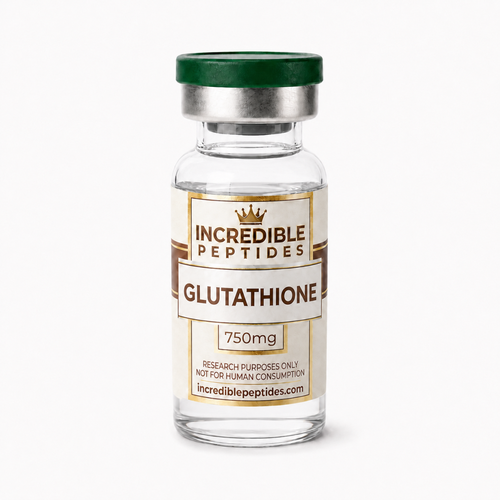
For analytical proof, use an HPLC method that separates GSH from GSSG. It is the most direct way to check whether a stock has drifted. Lot-specific storage conditions, identity and purity data for the glutathione research compound should be taken from the product listing. They should also be taken from its certificate of analysis.
Why a three-residue molecule repays careful handling
Glutathione is simple in chemistry but demanding in experiments. Its γ-glutamyl bond explains why normal peptidases leave it alone. Its cysteine thiol explains both its role in biology and its instability in solution. The two-to-one ratio in the GSH/GSSG couple explains why absolute amounts matter as much as ratios. Labs that control pH, metals, oxygen and sample handling get better data. Their data reflect the system under study, not the chemistry of the tube.
References
- Meister A, Anderson ME. Glutathione. Annual Review of Biochemistry, 1983.
- Schafer FQ, Buettner GR. Redox environment of the cell as viewed through the redox state of the glutathione disulfide/glutathione couple. Free Radical Biology and Medicine, 2001.
- Tietze F. Enzymic method for quantitative determination of nanogram amounts of total and oxidized glutathione. Analytical Biochemistry, 1969.
- Ellman GL. Tissue sulfhydryl groups. Archives of Biochemistry and Biophysics, 1959.
- Griffith OW, Meister A. Potent and specific inhibition of glutathione synthesis by buthionine sulfoximine (S-n-butyl homocysteine sulfoximine). Journal of Biological Chemistry, 1979.
Research Product in This Article
Educational content for research use only. Not for human consumption. Nothing here is medical advice or a claim about safety or efficacy.
 DO