Peptide Bonds and Amino Acid Sequences Explained
How amino acids link through peptide bonds, how sequences are written in one- and three-letter code, and why residue order defines a peptide.
 DODaniel Okafor, MSResearch Analyst, Molecular Biology~6 min read
 FundamentalsPhoto: Gannu03 / Wikimedia Commons
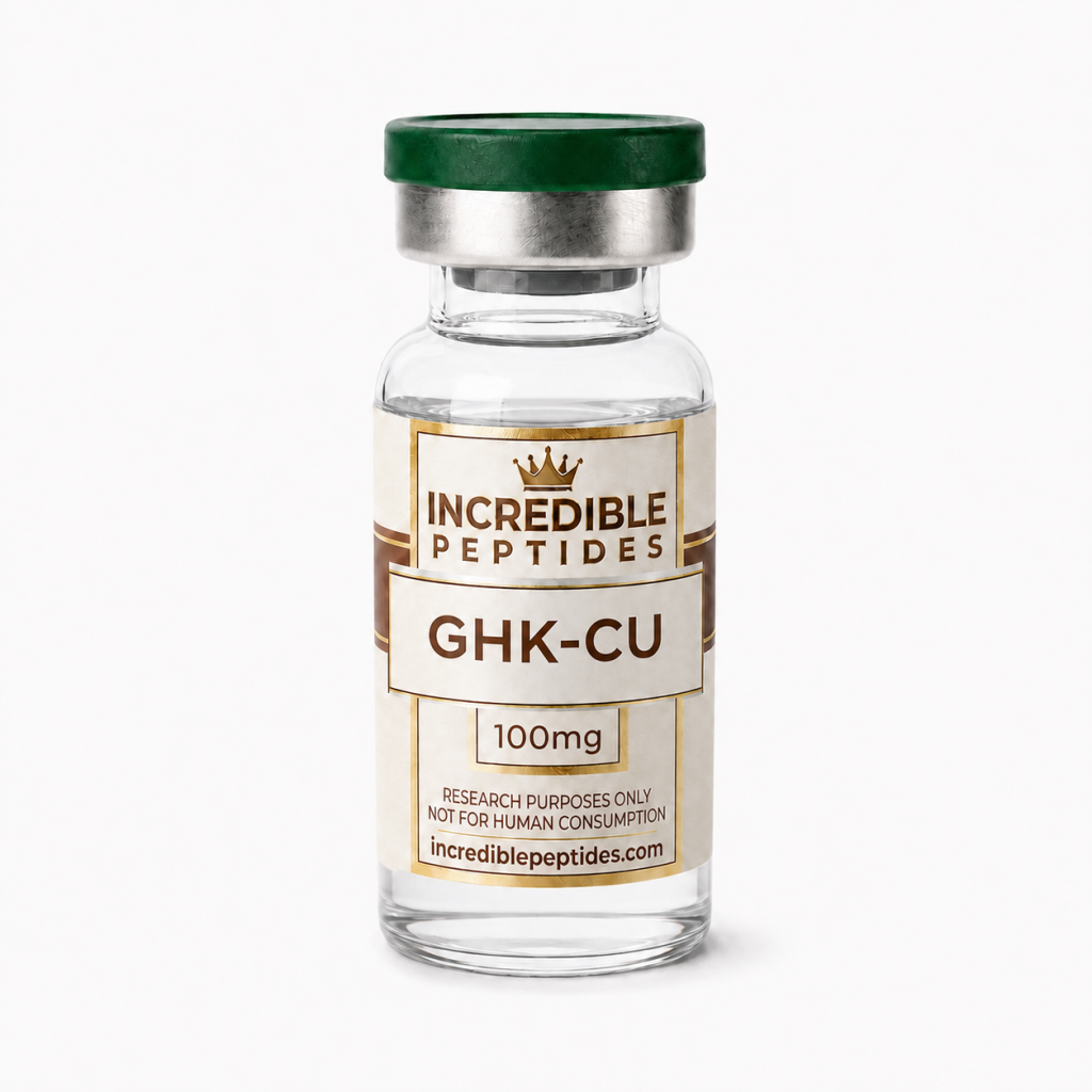
The peptide bond is the amide linkage that joins one amino acid to the next, and it is the most important covalent bond in peptide and protein chemistry. Every peptide, from a two-residue dipeptide to a long polypeptide, is a sequence of amino acid residues held together by these bonds, and the order of those residues is what distinguishes one molecule from another. Even product names are often sequences in disguise: GHK-Cu, a copper complex of the tripeptide Gly-His-Lys, takes its name directly from the one-letter codes of its three residues.
For students and researchers, a working command of peptide-bond chemistry and sequence notation is needed to read a specification, interpret a mass spectrum or recognize that two similarly named compounds are chemically distinct. This article covers how the bond forms and behaves, how sequences are written, what the termini signify and how common modifications are indicated.
The Peptide Bond
Formally, a peptide bond results from a condensation reaction between the α-carboxyl group of one amino acid and the α-amino group of another, with the loss of one molecule of water. The product is a secondary amide, –CO–NH–. In cells the reaction takes place on the ribosome, using aminoacyl-tRNAs as activated building blocks. In the laboratory the carboxyl component must likewise be chemically activated, because direct condensation of free amino acids in water is thermodynamically unfavourable; solid-phase peptide synthesis relies on coupling reagents for exactly this purpose.
Planarity and resonance
The peptide bond has partial double-bond character. The lone pair on the amide nitrogen is delocalized toward the carbonyl oxygen, so the C–N bond is shorter than a typical C–N single bond and rotation about it is restricted. As a result, the atoms of the peptide group (the carbonyl carbon and oxygen, the amide nitrogen and its hydrogen, and the two flanking α-carbons) lie approximately in one plane. Flexibility along the backbone comes instead from rotation about the single bonds on either side of each α-carbon, described by the dihedral angles phi (φ) and psi (ψ). Only certain combinations of φ and ψ avoid steric clashes, and those permitted regions are what make regular secondary structures such as α-helices and β-sheets possible.
Trans and cis configurations
A planar peptide bond can adopt a trans or a cis configuration. The trans form, with consecutive α-carbons on opposite sides of the bond, is strongly favoured for most residue pairs. The notable exception is a bond that precedes proline. Because proline’s side chain loops back onto its own backbone nitrogen, the energy difference between the two forms is smaller, and cis X-Pro bonds occur at measurable frequency. Slow cis-trans isomerization at proline can therefore matter when studying the conformation of proline-containing sequences.
Under neutral conditions the peptide bond is kinetically stable, but it can be hydrolyzed by acids, bases and proteolytic enzymes. Researchers handling peptides in solution should keep this in mind, along with side-chain degradation routes such as deamidation of asparagine and oxidation of methionine or cysteine.
Reading One- and Three-Letter Codes
The IUPAC-IUBMB Joint Commission on Biochemical Nomenclature defines standard abbreviations for the twenty proteinogenic amino acids. Each has a three-letter code (Gly, His, Lys, Pro, Val and so on) and a one-letter code (G, H, K, P, V). Three-letter codes are easier to read and are usually joined with hyphens, as in Gly-His-Lys. One-letter codes are more compact and are the norm for longer sequences and database entries, so the same tripeptide is written GHK.
Some one-letter codes are intuitive and some are not. G for glycine and H for histidine follow the first letter of the name, but K for lysine, W for tryptophan, F for phenylalanine and Y for tyrosine were assigned because the obvious letters were already taken. Confusing these is a common transcription error, particularly when sequences are copied by hand between documents. Unless stated otherwise, residues are assumed to be the L-enantiomers; D-amino acids are indicated with a D- prefix in three-letter notation or, in many one-letter conventions, a lower-case letter.
N-Terminus and C-Terminus
A linear peptide has direction. One end carries a free α-amino group, the N-terminus, and the other a free α-carboxyl group, the C-terminus. By convention sequences are written from N- to C-terminus, left to right, which mirrors the direction of ribosomal synthesis, and residues are numbered in the same direction.
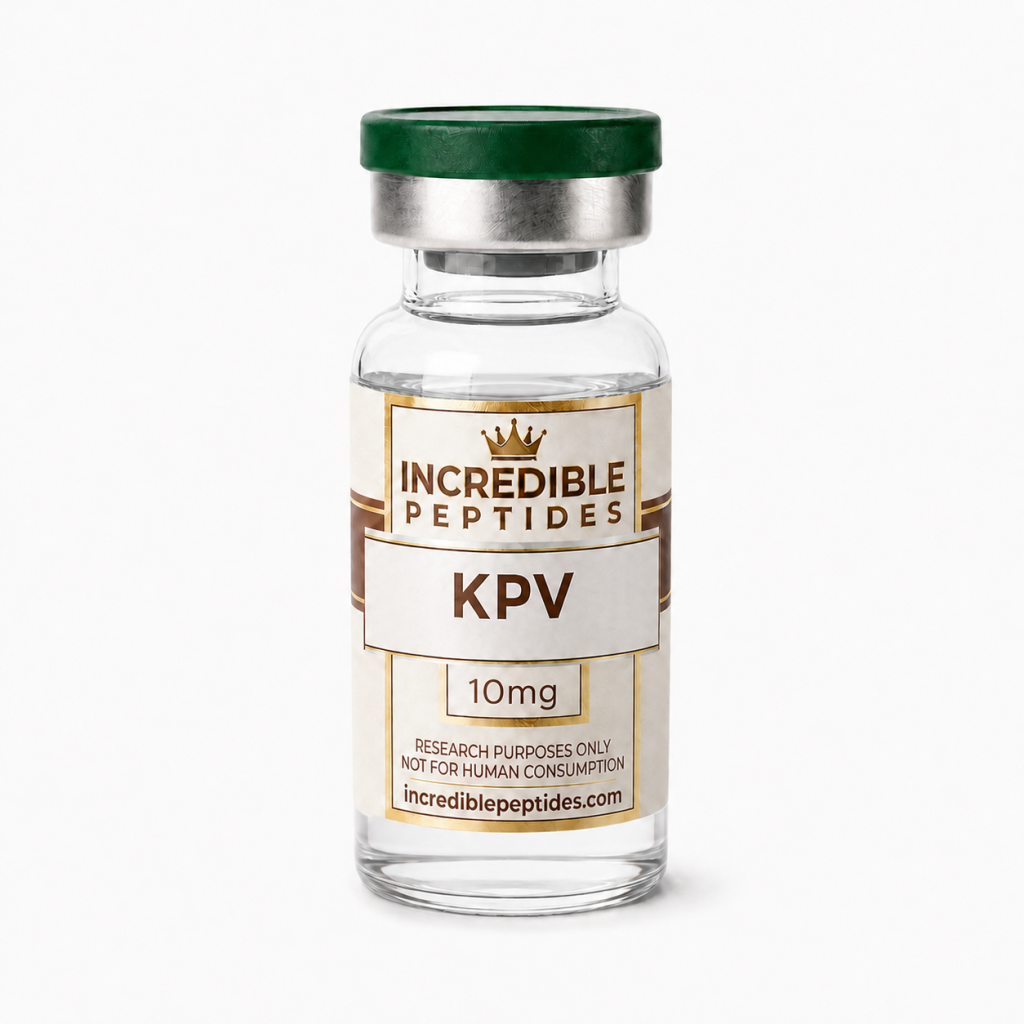
Direction matters because reversing a sequence produces a different molecule. Gly-His-Lys and Lys-His-Gly contain the same amino acids and have the same elemental composition and molecular mass, yet they present different groups at each end and place side chains at different positions along the backbone. A measurement of intact mass alone cannot tell them apart; fragmentation by tandem mass spectrometry or comparison with a reference standard is required. Consider KPV, the tripeptide Lys-Pro-Val: lysine sits at the N-terminus, valine at the C-terminus and proline between them. Its Lys–Pro bond is an example of the X-Pro linkage in which both cis and trans forms are accessible. The sequence corresponds to residues 11 to 13 at the C-terminal end of α-melanocyte-stimulating hormone (α-MSH), which illustrates how short research peptides are often defined as fragments of longer native sequences.
At neutral pH the free N-terminal amine is largely protonated and the free C-terminal carboxyl largely deprotonated, so the termini contribute charges in addition to those of ionizable side chains. That is one reason terminal modifications change a peptide’s physicochemical behaviour.
Modifications: Amidation, Acetylation and Metal Complexes
Many peptides differ from the plain linear sequence through defined modifications, and the notation must capture them. C-terminal amidation replaces the terminal –COOH with –CONH2, removing a negative charge; it is written by appending –NH2 to the sequence, as in …-Pro-Val-NH2. N-terminal acetylation caps the amino terminus with an acetyl group and is written with the prefix Ac-. Native α-MSH, for instance, is both N-acetylated and C-terminally amidated. A free acid and its amide are distinct compounds whose monoisotopic masses differ by just under 1 Da, so a specification should state which form is meant.
Other common modifications include disulfide bridges between cysteine residues, phosphorylation of serine, threonine or tyrosine, pyroglutamate formation from an N-terminal glutamine, and the incorporation of D-amino acids or non-proteinogenic residues. Metal complexes form a separate category, because the metal is coordinated rather than covalently built into the backbone. The copper(II) complex of glycyl-L-histidyl-L-lysine, GHK-Cu, is a standard example: the suffix in GHK-Cu identifies the bound metal, and characterizing such a complex means considering both the peptide ligand and its metal content.
Worked Examples from the Catalog
Putting these conventions together, consider how two short catalog sequences decode. GHK, written out as Gly-His-Lys, has glycine at the N-terminus, the smallest amino acid with only a hydrogen atom as its side chain; histidine in the middle, whose imidazole ring is partly protonated near neutral pH and can coordinate metal ions; and lysine at the C-terminus, carrying a positively charged ε-amino group. The molecule contains two peptide bonds, Gly–His and His–Lys.
KPV reads Lys-Pro-Val. The N-terminal lysine supplies both an α-amino and an ε-amino group, proline is a cyclic residue whose ring constrains the backbone, and valine provides a branched hydrophobic side chain at the C-terminus. Again there are two peptide bonds; the first, Lys–Pro, is the one that precedes proline. In both examples, rearranging the letters would create a different chemical entity even though the composition is unchanged. That point underlies structure-activity work, in which individual residues are substituted or reordered to test their contribution, as discussed in our article on peptide structure-activity relationships.
Why Sequence Literacy Matters
The peptide bond gives the backbone its planarity and its direction, and sequence notation turns that chemistry into a compact, unambiguous record. Reading a sequence correctly, including its termini and any modifications, is the first step in checking a specification, predicting a molecular mass or designing a scrambled-sequence control. Readers who want to see what changes as chains grow longer and begin to fold can continue with our comparison of peptides vs proteins.
References
- IUPAC-IUBMB Joint Commission on Biochemical Nomenclature. Nomenclature and Symbolism for Amino Acids and Peptides (Recommendations 1983). Pure and Applied Chemistry, 1984.
- Nelson DL, Cox MM. Lehninger Principles of Biochemistry. W. H. Freeman.
- Berg JM, Tymoczko JL, Gatto GJ, Stryer L. Biochemistry. W. H. Freeman.
- Merrifield RB. Solid phase peptide synthesis. I. The synthesis of a tetrapeptide. J. Am. Chem. Soc. 1963, 85, 2149.
Research Products in This Article
Educational content for research use only. Not for human consumption. Nothing here is medical advice or a claim about safety or efficacy.