What Is GLP-1? Glucagon-Like Peptide-1 Biology for Researchers
Glucagon-like peptide-1 from gene to receptor: proglucagon processing, GLP-1R signalling, why the native peptide is short-lived and how analogues fix that.
 DODaniel Okafor, MSResearch Analyst, Molecular Biology~6 min read
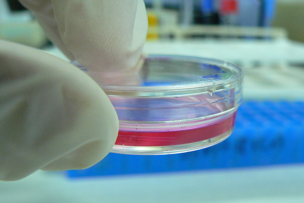 MechanismsPhoto: kaibara87 / Wikimedia Commons, CC BY 2.0
Glucagon-like peptide-1 is a short peptide hormone produced when the proglucagon precursor is cut by a specific processing enzyme in specialised gut cells. For a laboratory scientist, the question of what is GLP-1 has a precise biochemical answer: a 30- or 31-residue peptide that activates one class B1 G protein-coupled receptor and is inactivated within minutes by a widely distributed protease. Those three facts explain most of what makes the molecule interesting to study, and they explain why so much medicinal chemistry has gone into building longer-lived analogues.
This article walks through proglucagon processing, the active forms of GLP-1, the cells that make it, the GLP-1 receptor and its signalling, enzymatic degradation, and the structural strategies behind stabilised analogues such as semaglutide, which is supplied for laboratory work as a GLP-1 class research peptide.
One gene, several peptides: proglucagon processing
The proglucagon gene (GCG) encodes a precursor of roughly 160 amino acids that carries several hormone sequences in tandem. Which peptides a cell releases depends on the prohormone convertase it expresses. In pancreatic alpha cells, prohormone convertase 2 (PC2) predominates, and the main products are glucagon, glicentin-related pancreatic polypeptide and the major proglucagon fragment. In intestinal L-cells, prohormone convertase 1/3 (PC1/3) cleaves at different sites and liberates glicentin, oxyntomodulin, GLP-1, intervening peptide-2 and GLP-2. A population of preproglucagon neurons in the nucleus of the solitary tract in the brainstem follows the intestinal pattern and also produces GLP-1.
The name reflects history rather than function. When the proglucagon sequence was deduced from cloned cDNA in the early 1980s, two segments downstream of glucagon showed obvious sequence similarity to it and were labelled glucagon-like peptides 1 and 2. GLP-1, glucagon, GLP-2 and the related gut hormone GIP (gastric inhibitory polypeptide) form a structurally related family, which is why their receptors also resemble one another.
The active forms: GLP-1(7-37) and GLP-1(7-36)amide
The first full-length sequence predicted from the gene, GLP-1(1-37), proved only weakly active. The biologically important forms start at histidine 7 of that original numbering, after processing removes the first six residues. Two active species exist. GLP-1(7-37) ends in glycine, while in GLP-1(7-36)amide that glycine has served as the nitrogen donor for C-terminal amidation of arginine 36 by peptidylglycine alpha-amidating monooxygenase. The two are generally regarded as similarly active at the receptor. Researchers keep the 1-37 numbering by convention, so the N-terminal residue is always called His7.
| Position | Native residue | Why it matters |
|---|---|---|
| 7 | His | N-terminal residue; essential for receptor activation |
| 8 | Ala | Second residue; defines the DPP-4 cleavage site |
| 26 | Lys | Side chain used as the lipidation site in several analogues |
| 34 | Lys | Second lysine; often replaced by Arg to leave a single acylation site |
| 36/37 | Arg-NH2 or Arg-Gly | C-terminus of the amidated or glycine-extended form |
The full sequence of GLP-1(7-37) in one-letter code is HAEGTFTSDVSSYLEGQAAKEFIAWLVKGRG. In solution the peptide is largely unstructured, but in membrane-mimetic environments and when bound to its receptor the segment from roughly residue 13 to the C-terminus adopts an alpha-helix.
L-cells and the secretion context
L-cells are open-type enteroendocrine cells whose apical surface faces the gut lumen, letting them sense luminal contents directly. They become more numerous toward the distal ileum and colon, and many co-store peptide YY. In preclinical work, secretion has been linked to cell-surface sensors such as the bile acid receptor TGR5 and GPR119. Common in vitro models include the murine GLUTag and STC-1 lines and primary rodent intestinal cultures, each an imperfect stand-in for native L-cells.
The GLP-1 receptor and its signalling
GLP-1 acts through the GLP-1 receptor (GLP-1R), cloned from a rat pancreatic islet library by Thorens in 1992. It belongs to class B1 of the G protein-coupled receptor superfamily, the same family as the glucagon, GIP, secretin and calcitonin receptors. Class B1 receptors share a large N-terminal extracellular domain stabilised by three conserved disulfide bonds, attached to the seven-helix transmembrane core. Ligands for this receptor family, from native-sequence hormones to engineered analogues, sit among the single-compound research peptides used in receptor pharmacology.
Peptide recognition follows a two-domain model. The C-terminal helical portion of GLP-1 docks into a groove on the extracellular domain, which supplies much of the binding affinity. The N-terminal region, including His7, then reaches into the transmembrane bundle, where it engages residues that stabilise the active conformation. Cryo-electron microscopy structures of GLP-1R bound to agonist and the Gs heterotrimer, first reported in 2017, made this arrangement visible at near-atomic resolution and have since guided analogue design.
The dominant signalling pathway is Gαs coupling, which activates adenylyl cyclase and raises intracellular cAMP; downstream effectors include protein kinase A and Epac2. The receptor also recruits β-arrestins, undergoes agonist-driven internalisation and can activate ERK1/2 and calcium signalling depending on cell type. Two natural tools are worth knowing. Exendin-4, a 39-residue peptide isolated from Gila monster (Heloderma suspectum) venom, is a potent GLP-1R agonist that resists DPP-4, and its truncated form exendin(9-39) is a widely used competitive antagonist. The broader principles are covered in our primer on peptide receptor binding.
Why native GLP-1 is so short-lived
The main reason is dipeptidyl peptidase-4 (DPP-4), a serine protease found on cell surfaces and in soluble form in plasma. DPP-4 removes N-terminal dipeptides from substrates that carry proline or alanine in the second position. GLP-1 fits that pattern exactly, and cleavage of the His7-Ala8 dipeptide yields GLP-1(9-36)amide, which binds the receptor far more weakly and has little agonist activity. Neutral endopeptidase 24.11 cleaves the peptide at internal sites, and renal filtration clears the small molecule rapidly. Together these routes give intact GLP-1 a circulating half-life of only a few minutes in mammalian studies.
This has practical consequences at the bench. Serum and plasma contain active DPP-4, so incubating native GLP-1 in biological matrices without an inhibitor produces a mixture of intact and truncated peptide, and preclinical sampling protocols routinely add a DPP-4 inhibitor to collection tubes. Our article on peptidases covers the enzyme classes involved in more detail.
Engineering longer-lived analogues
Two complementary strategies dominate. The first protects the N-terminus. Exendin-4 has glycine at position 2 and is a poor DPP-4 substrate, which pointed the way. In synthetic analogues, Ala8 is often replaced with α-aminoisobutyric acid (Aib), a non-coded residue whose two methyl groups on the alpha carbon block recognition by the enzyme while preserving receptor activation.
The second strategy attaches a fatty acid to a lysine side chain so that the peptide binds reversibly to serum albumin. Albumin-bound peptide escapes renal filtration and is partly shielded from proteases, and the lipid can also promote self-association. Semaglutide combines both ideas: Aib at position 8, arginine replacing Lys34, and Lys26 carrying a linker of two short oligo(ethylene glycol) units and a γ-glutamic acid capped with an 18-carbon fatty diacid. The GLP-1 (semaglutide) research peptide is therefore a lipidated analogue rather than the native hormone, a distinction that matters when choosing a reference agonist for receptor assays. The structural details are covered in our semaglutide peptide structure profile.
Bringing the biology together for lab work
GLP-1 is a product of tissue-specific precursor processing, recognised by a class B1 receptor through a two-step binding mechanism and switched off by DPP-4 within minutes. Analogue chemistry answers each vulnerability in turn. When designing experiments, decide early whether the question concerns the native peptide or an engineered analogue, control for protease activity and albumin in the assay buffer, and confirm identity and purity against the lot certificate of analysis. All compounds discussed are intended for laboratory research use only and are not for human or veterinary use.
References
- Holst JJ. The physiology of glucagon-like peptide 1. Physiological Reviews. 2007;87(4):1409–1439.
- Thorens B. Expression cloning of the pancreatic beta cell receptor for glucagon-like peptide 1. Proceedings of the National Academy of Sciences USA. 1992;89(18):8641–8645.
- Eng J, Kleinman WA, Singh L, Singh G, Raufman JP. Isolation and characterization of exendin-4, an exendin-3 analogue, from Heloderma suspectum venom. Journal of Biological Chemistry. 1992;267(11):7402–7405.
- Mentlein R, Gallwitz B, Schmidt WE. European Journal of Biochemistry. 1993;214(3):829–835.
- Zhang Y, Sun B, Feng D, et al. Cryo-EM structure of the activated GLP-1 receptor in complex with a G protein. Nature. 2017;546:248–253.
- Lau J, Bloch P, Schäffer L, et al. Journal of Medicinal Chemistry. 2015;58(18):7370–7380.
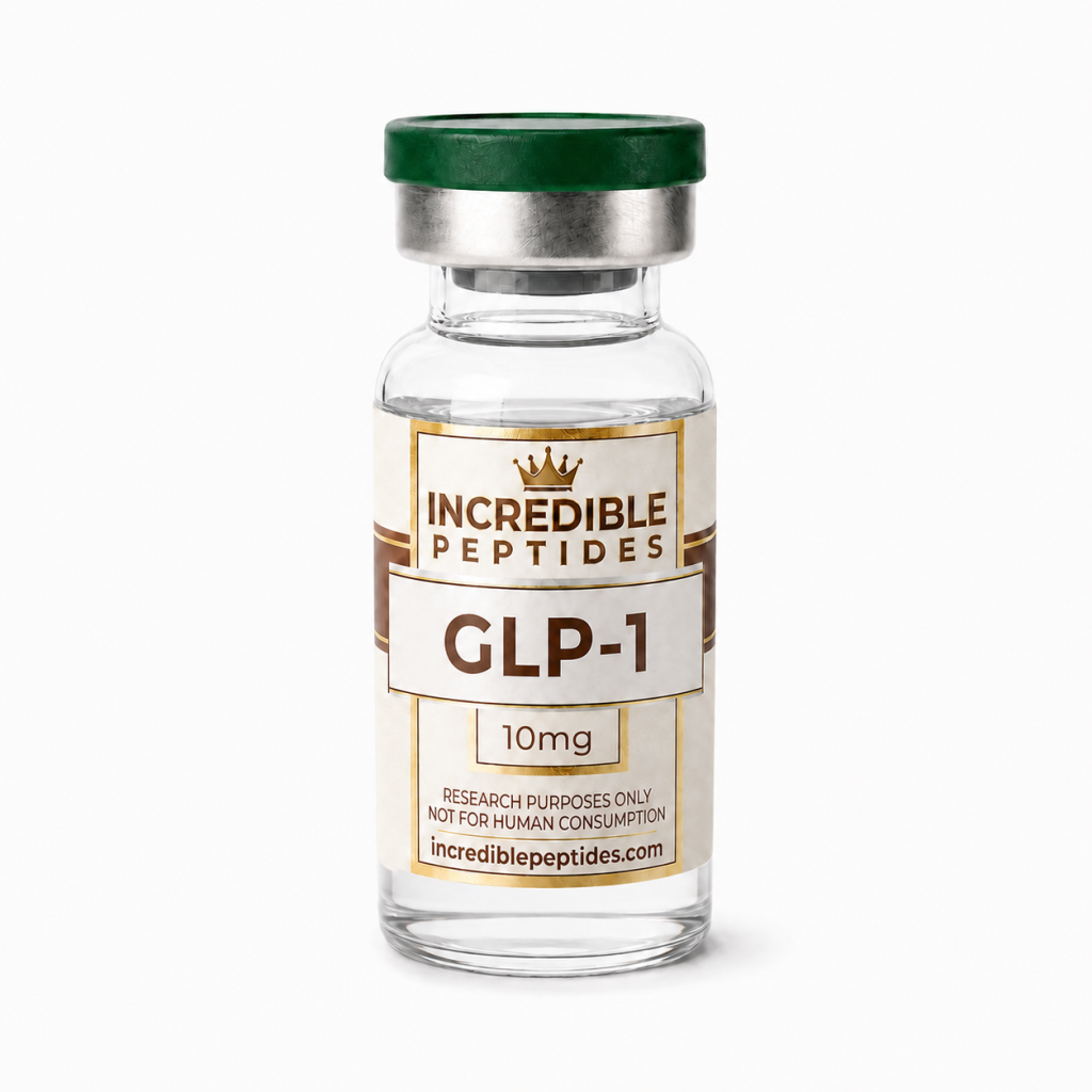
Research Product in This Article
Educational content for research use only. Not for human consumption. Nothing here is medical advice or a claim about safety or efficacy.