
Retatrutide (RETA-3): A Triple Receptor Agonist Research Peptide
How retatrutide is built, why it activates three related class B1 receptors, and what in vitro receptor profiling can and cannot tell you.
 DODaniel Okafor, MSResearch Analyst, Molecular Biology~4 min read
 Compounds
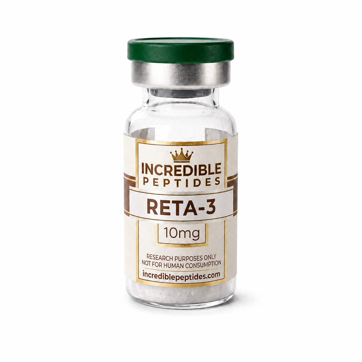
Retatrutide is a synthetic peptide engineered to activate three related receptors at once: the receptors for glucose-dependent insulinotropic polypeptide (GIPR), glucagon-like peptide-1 (GLP-1R) and glucagon (GCGR). That triple activity makes the retatrutide peptide a useful tool for studying how a single ligand can be tuned across a family of closely related G protein-coupled receptors. In the research literature it appears under its development code, LY3437943, and in the research-peptide market it is sometimes called a “GLP-3” peptide, a nickname rather than the name of any natural hormone.
This article covers how retatrutide is built, why one sequence can engage three receptors, how that activity is measured in cells, and what laboratories should check when they work with RETA-3, our retatrutide research peptide. It stays at the level of chemistry and receptor pharmacology. For laboratory research use only; not for human or veterinary use.
Three Receptors From One Hormone Family
GIP, GLP-1 and glucagon are peptide hormones of similar length that share a common ancestry and a common receptor architecture. Each binds a class B1 GPCR made of a large extracellular domain that grips the ligand’s C-terminal helix and a transmembrane core that receives the ligand’s N-terminus. The N-terminal residues are what switch the receptor on, so small sequence changes in that region shift which receptors a peptide activates and how strongly. Our primer on peptide-receptor binding walks through this two-domain model in more detail.
Because the three hormones overlap in sequence, medicinal chemists have long built hybrid peptides that borrow residues from more than one of them. Dual GIPR/GLP-1R agonists came first; retatrutide extends the idea by adding glucagon receptor activity, giving a so-called triple agonist.
How Retatrutide Is Built
A GIP-based backbone
Retatrutide is a peptide of roughly 39 residues whose sequence is derived largely from GIP, with substitutions chosen to bring in GLP-1R and GCGR activity. It includes non-proteinogenic residues such as α-aminoisobutyric acid (Aib), which restrict backbone flexibility and protect the N-terminal region from dipeptidyl peptidase-4, the enzyme that rapidly truncates native GLP-1 and GIP. The exact published sequence should be taken from the primary literature or the lot certificate rather than from secondary sources.
Lipidation and albumin binding
Like several modern incretin-class research peptides, retatrutide carries a long-chain fatty diacid attached through a hydrophilic linker to a lysine side chain. The lipid moiety binds serum albumin non-covalently, which greatly slows clearance in animal models. For the bench scientist the practical consequences are different: the lipid makes the molecule amphiphilic, more prone to adsorb to plastics, and sensitive to the albumin or serum content of assay buffers, all of which can shift apparent potency.
Measuring Activity at Each Receptor
All three receptors couple mainly to Gs, so the standard readout is cAMP accumulation in cell lines that express one receptor at a time. Running the same peptide through GIPR, GLP-1R and GCGR cell lines side by side gives three concentration-response curves, and comparing their EC50 values describes the compound’s balance across the receptors. Our article on cAMP and other cell signaling assays explains how these assays are set up and read.
Several cautions apply. Potency depends on receptor expression level, so a cell line with high receptor density can make a weak agonist look strong. Species matters: rodent and other receptor orthologues can respond differently. And serum albumin in the buffer binds lipidated peptides, so the same compound can appear less potent in albumin-containing medium than in albumin-free buffer. Published comparisons only make sense when these conditions match, a point covered in our guide to binding affinity, IC50 and EC50. Researchers comparing retatrutide with single and dual agonists often keep reference ligands for each receptor in the same plate, and they may also screen other receptor-active research peptides in the catalog as comparators.
Beyond cAMP: Bias and Receptor Trafficking
A ligand can activate cAMP signaling strongly while recruiting β-arrestin weakly, or the reverse. This signaling bias, along with differences in how quickly each receptor is internalized and recycled, is an active research area for multi-receptor peptides. Arrestin recruitment assays, receptor internalization imaging and phosphorylated ERK readouts add information that a single cAMP curve cannot. For a triple agonist, bias can differ at each of the three receptors, which is one reason these compounds are studied receptor by receptor rather than as a single number.
Preclinical Context
Published characterization of LY3437943 includes in vitro receptor profiling and studies in rodent models of metabolic signaling. Those animal findings are preclinical and describe the models studied; they are not evidence about anything outside those systems.
Handling, Identity and Purity
Retatrutide is typically supplied as a lyophilized powder. Its size and lipidation make it more prone to aggregation and surface adsorption than short unmodified peptides, so low-binding tubes, freshly prepared stock solutions and minimal freeze-thaw cycling are sensible defaults. Mass spectrometry should show the expected intact mass including the lipid side chain, and reversed-phase HPLC typically elutes the lipidated peptide late with a broadened peak if aggregation is present. The guide to reading a peptide COA explains what to check on the certificate.
Why the Triple-Agonist Design Matters for Research
Retatrutide is a clear example of how a peptide can be engineered across a receptor family: start from one hormone’s backbone, borrow residues from its relatives, add residues that resist proteolysis, and attach a lipid for albumin binding. Each of those steps can be probed in the lab with receptor-specific cell lines, matched assay conditions and careful analytical work on lot-matched RETA-3 material. Treated that way, it is a useful model compound for anyone studying multi-receptor pharmacology in class B1 GPCRs.
References
- Coskun T, et al. Cell Metabolism, 2022 (characterization of LY3437943 as a GIP, GLP-1 and glucagon receptor agonist).
- Hollenstein K, et al. Insights into the structure of class B GPCRs. Trends in Pharmacological Sciences, 2014.
- Mentlein R. Dipeptidyl-peptidase IV (CD26): role in the inactivation of regulatory peptides. Regulatory Peptides, 1999.
- Alexander SPH, et al. The Concise Guide to Pharmacology: G protein-coupled receptors. British Journal of Pharmacology (IUPHAR/BPS).
Research Product in This Article
Educational content for research use only. Not for human consumption. Nothing here is medical advice or a claim about safety or efficacy.